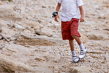
Foot and Ankle Doctor Blog Space

Don’t Be Flip-Flopping Around This Summer

At Foot & Ankle Doctors, Inc. we understand that flip-flops are a summer staple, and have become so popular that some companies have created flip-flops with a built in bottle opener. Although flip-flops and sandals are comfortable and fashionable, they aren’t the best footwear for you.

Flip-flops have little to no arch support, they have minimal cushioning and shock absorption and can lead to foot pain if worn all day long. Common foot injuries associated with flip-flops include: blisters, stress fracture of the metatarsals, plantar fasciitis, ankle sprains and tendonitis. It’s okay to wear flip-flops when running a few errands, at the beach, or at a public pool, but they should not be worn when walking long distances, doing yard work and playing sports.

Patients with diabetes should avoid flip-flops because they expose your feet and increase your chances of lacerations and puncture wounds leading to serious infections.

What to look for in a summer sandal:

  • High quality material such as soft leather that will not irritate the skin
  • A sturdy pair that will not fold in half when bent
  • Built in arch support

It’s important to remember that the sun can easily damage the skin on the feet and sunscreen should be applied to protect them from sun damage

Dr. Nejad

Athlete’s Foot

Athlete’s foot can be an embarrassing problem, despite its name anyone can contract athlete’s foot. It is caused by fungal infections and is easily contracted when wearing damp socks in tight shoes or walking barefoot in public locker rooms and shower areas.

Symptoms include cracked, scaling, itchy red skin between the toes and on the bottom on feet. In some cases blisters or ulcers may form. Athlete’s foot is contagious and it is best to avoid scratching or picking the affected areas on the feet.

Over the counter medication can be used to treat Athlete’s foot, however if your symptoms do not get better in a few weeks come see us at Foot & Ankle Doctors, Inc. If we cannot diagnose your symptoms classically as Athlete’s Foot a small shave biopsy will be taken and sent to the lab to be examined under a microscope before topical or oral anti-fungual medications will be dispensed.

  • To avoid Athlete’s Foot:
  • Keep your feet dry
  • Change your socks regularly
  • Don’t walk around barefoot in public places
  • Wear well ventilated shoes

OATS for your Ankle Pain

Young adolescents complaining of ankle pain, joint popping or locking with decreased range of motion of the ankle and swelling and tenderness are signs of osteochondritis dissecans (OCD). OCD most often occurs in athletic males between the ages of 10 and 20. A small piece of cartilage becomes loose and can detach in some cases. If the fragment of cartilage migrates and gets stuck in the ankle joint, surgical intervention is required. OCD can increase your chances of developing osteoarthritis of the ankle joint.

Imaging

Physicians at Foot & Ankle Doctors, Inc. will take x-rays of your ankle and stage the severity of the injury from 1 to 4.

Treatment

Treat of early stages of OCD involve immobilization of the ankle joint in a non-weight bearing cast to avoid stress on the ankle. Physical therapy is important to increase range of motion and strengthen muscles that support the ankle joint. If conservative treatments fail after 3 months or a fragment of bone is caught in the ankle joint, surgery is required.Surgical intervention includes arthroscopic treatment to clean the ankle joint of debris. Osteoarticular Transfer System (OATS) surgery is another treatment that replaces damaged cartilage relieving pain and resorting movement. Small plugs of healthy cartilage are taken from another area of the ankle joint and transferred to damaged area. Patients will be on crutches for 6 to 12 weeks after surgery and long term physical therapy is necessary to restore range of motion and relieve pain.

For more information visit us at Foot & Ankle Doctors, Inc.

Dr. Nejad

Bone Spurs in the Big Toenail

If you notice a soft pink mass on the tip of your big toe or sensitive area under the nail plate that is protruding and is painful when pressure is applied you may have a subungal exostosis. A subungual exostosis is a benign bone growth that arises from the dorsal surface of the distal phalanx (a small bone at the end of the toe) and causes pain and deformity of the nail.

Common causes for subungual exostosis include trauma, narrow shoes, previous nail surgery and certain biomechanical deformities such as a Hallux limitus.

At Foot & Ankle Doctors, Inc. x-rays of the foot will be taken to examine the bony growth and its involvement of the big toe. Differential diagnosis include: osteochondroma, Nora’s lesion, and osteosarcoma.

Conservative treatment includes purchasing a wider shoe and continuing with routine podiatric care.

Surgical removal is the recommended course of action. There are various surgical approaches and incisions that can be used to remove the bony prominence depending on its severity. In most cases the big toenail is removed to gain access to the bony prominence, which is then cut out. A surgical shoe will be dispensed and should be worn for 1-2 weeks.

Dr. Dardashti

Puncture Wound

Stepping on a thumbtack, nail or piece of glass is something that shouldn’t be taken lightly. Most people try to treat themselves at home, however any object that penetrates the skin can lead to an infection especially if the object is not clean.

A puncture wound can lead to cellulitis, an infection caused by streptococcus or staphylococcus bacteria. Cellulitis will cause swelling and redness to the skin. The infected area will be warm and painful to the touch and you may develop a fever. If the bacteria enter the bloodstream the infection can spread to the rest of the body and sometimes lead to a life threatening infection know as necrotizing fasciitis.

If you step on a nail, seek medical attention immediately. If your tetanus shots are not up to date, your physician will administer a tetanus shot to protect you against lockjaw, an infection that can cause your muscles to painfully contract.

Treatment
Come see us at the Foot & Ankle Doctors, Inc
. within 24 hours after injury. We will remove the foreign object and take x-rays to ensure no pieces remain in the foot. We will clean the wound and monitor your progress during healing. In some cases antibiotics may be prescribed.

Complications
Infection is always a concern after a puncture wound. Signs of infection include warmth, redness, swelling, soreness and sometimes drainage. If the object penetrated to bone, a bone infection may occur. In some cases a painful scar or hard cyst may develop.

Dr. Farshid Nejad

Acute Compartment Syndrome in the Foot

Compartment syndrome is a painful condition that occurs from an increase in pressure within the muscles. Increase in pressure above normal levels can decrease blood supply to the muscles and nerves in that area which can result in permanent tissue damage.

Acute compartment syndrome is a medical emergency. It is usually caused by trauma such as a crush injury, a fracture or a constricting bandage or cast.

Chronic compartment syndrome is not a medical emergency. It is usually caused by repetitive physical activity such as running.

There are 9 compartments in the foot and each compartment has a fascia covering to keep the tissues in place. This fascia does not stretch easily and following an injury, swelling or bleeding can occur, resulting in increased pressure within the compartments. If the pressure is not released, permanent tissue damage can occur.

Symptoms – The 6 P’s of Compartment Syndrome

  • Pain out of proportion and not relieved by narcotics
  • Pulselessness
  • Parasthesia
  • Paralysis
  • Pallor
  • Poikilothermia

If you experience these symptoms, seek medical advice immediately

Diagnosis
A wicker catheter is a device that is used to measure compartment pressures. Normal compartment pressure in the foot is between 0-8mmHg. A compartment pressure measuring >30mmHg or 10-30mmHg below diastolic pressure is concerning and emergent fasciotomy may be needed.

Treatment
Depending on which foot compartments are affected incisions are made over the compartments to relive pressure. Fasciotomy incisions are left open 5 days to allow edema and swelling to dissipate. After the pressure has been adequately reduced the surgical sites are closed.

Acute compartment syndrome is a surgical emergency and if not treated immediately permanent damage and tissue death can occur. For more information on acute compartment syndrome come see one of our physicians at Foot & Ankle Doctors, Inc

Dr. Dardashti

Fracture Healing

All broken bones in the body undergo a natural healing process. Bone healing is a slow process and can take up to 8 weeks. Immobilization and compression is necessary for optimal bone healing.

There are four phases of bone healing

  • Inflammation: occurs within the first 48 hours. Swelling can help provide stability
  • Soft callus: occurs during the first two months. Fibrous tissue and cartilage tissue develop at the ends of the fracture site
  • Hard Callus: occurs during the 3rd and 4th month. The soft callus is replaced with bone and is visible on x-ray
  • Bone remodeling: Lasts for several months. The bone continues to return to its normal shape. Bone can regenerate back to 100% of its strength after a fracture

At Foot & Ankle Doctors, Inc we like to immobilize patients with fractures. Immobilization is an important part in promoting bone healing. Depending on the type of fracture a surgical shoe or boot may be used. In more severe cases surgery may be needed to fixate the fracture site with screws, plates or wires. A cast will be applied afterwards and physical therapy recommended once the bone is adequately healed.

Factors that Impair Bone Healing

  • Early weight bearing
  • Smoking
  • Fracture ends greater than 1cm apart
  • Poor nutrition
  • Certain medications

If bone healing is slow or delayed, our physicians can provide a variety of treatments to enhance bone growth such as bone stimulators, use of bone growth proteins and bone grafts.

Dr. Dardashti

Emergent Gas Gangrene

Gas gangrene is an emergent case where a bacterium, most commonly Clostridium perfringens grows inside the body and produces harmful toxins and gas causing tissue death. It can be caused by other bacteria such as Group A streptococcus, Staphylococcus aureus and Vibro vulnificus.

Gas gangrene most often occurs at a wound site, recent surgical site or trauma site. It can develop suddenly and rapidly accelerate causing irreversible tissue damage.

Symptoms of Gas Gangrene

 Change in color of the skin to a pale to brownish-red color


 Crepitus– gas can be felt as a cracking sensation when pressing on the swollen area


 Brown fluid filled blisters


 Fever


 Sweating

If not treated immediately a person can go into shock which will lead to kidney failure and eventually lead to death.

Diagnosis

Gas Gangrene can be diagnosed by clinical examination. To confirm the presence of Clostridium bacteria a tissue culture may be taken. Blood cultures can also be used to determine a bacteria infection. X-rays can be used to identify the presence of gas in the soft tissue.

Treatment

Gas gangrene is an emergent case and surgical intervention is required. Surgical debridement of the damaged and infected tissue must be removed. In severe cases partial amputation may be required to prevent further spread of the infection. IV antibiotics will be prescribed. In some cases hyperbaric oxygen (HBO) treatment may be implemented. Clostridium bacteria cannot survive in an oxygen rich environment and therefore HBO can be used to kill the bacteria.

Dr. Farshid Nejad

Pediatric Clubfoot

Every 1 in 1,000 births a child is born with a clubfoot, which can be devastating to parents. Pediatric clubfoot is curable and if the deformity is corrected early, children born with clubfoot are indistinguishable from other children with normal feet.


What is Clubfoot?


Clubfoot occurs when a child’s foot is turned inwards and the heel points downwards. In 50% of cases the deformity can occur in both feet. The calf muscles of the affected side are smaller than normal. Clubfoot is a congential deformity, meaning the child is born with the condition.


Causes


 Clubfoot is most often idiopathic (no apparent cause)


 Associated with genetic syndromes and birth defects


 Result from a neuromuscular disorder


Treatment


The “Ponseti” casting method is the preferred method for treating pediatric clubfoot. Treatment should begin within the first month of life for best results. The child’s foot is manipulated into the correct position through a serious of casting. 6-8 leg casts are applied every 5-7 days to slowly correct the deformity overtime. In some cases a small cut may be made to the heel cord to release contracture of the Achilles tendon. After the casting series is completed a special brace is required to be worn during naps and at night until the age of 4.


Clubfoot is visible at birth and can be detected by fetal ultrasound. If your child has been diagnosed with clubfoot come see us at Foot& Ankle Doctors, Inc for more information.


Dr. Dardashti

Neuropathic Joints

Charcot is a condition in patients who have neuropathy or loss of sensation. Charcot can lead to fractures and dislocations in the foot and ankle. As the disease progresses the joints collapse and the foot can take an abnormal shape known as a rocker-bottom foot. Diabetes is the most common cause of Charcot because diabetes is associated with poor blood flow, causing weakening of bones, and neuropathy, which is the main cause of Charcot. Other causes and risk factors include repetitive microtrauma and patients with peripheral neuropathy secondary to:

– Chronic alcoholism

– Leprosy

– Syphilis

– Poliomyelitis

Symptoms

There are various stages of Charcot and symptoms will vary depending on the stage of the deformity.

Stage 1: Red, hot, swollen foot/ankle.

X-rays show soft tissue edema and bony fragments

Stage 2: Decreased local edema foot/ankle

X-rays reveal coalesence of bony fragments

Stage 3: No local edema

X-rays show consolidation and remodeling of the bone

Imaging

In addition to reviewing your overall health, symptoms and discussing your medical history, our physicians at Foot& Ankle Doctors, Inc may order various imaging tests for further detailed pictures of your foot or ankle.

X-rays:

In early stages of Charcot, x-rays may appear normal. As the disease progresses the bones may fracture or dislocate. As a result repeat x-rays may be taken several weeks later.

MRI:

Ordered to evaluate soft tissue and when a bone infection is suspected

Bone Scans:

Helpful to differentiate between Charcot and infection. Charcot and an infection are positive on a technetium bone scan, however infection will only produce a hot spot on an indium bone scan and Charcot will not.

Non-Surgical Treatment

– Immobilization is important in the early stages of Charcot to protect the bones

from stress and shear forces that can further damage the bones

– Total contact cast changed every 1-2 weeks for 2-4 months

– Charcot restraint orthotic walker (CROW) boot

Custom Shoes designed to decrease the risk of ulcers

– Rocker bottom shoes will help reduce the risk of ulceration

Surgical Treatment

Depending on the type and extent of the deformity surgery may be recommended

Dr. Dardashti

Make an Appointment Same Day Appointments Available

BOOK NOW